


Preoperative Defense Against Complications
From hemostasis to nerve protection, predictable tooth extraction begins before the first instrument touches the tooth.
Chapters 16–25 of Extraction Techniques systematically address systemic complications, local complications and iatrogenic incidents associated with tooth extraction.
One principle runs throughout the discussion: many extraction complications can be reduced through appropriate preoperative assessment, standardized intraoperative technique and structured postoperative care.
Prevention Over Rescue
Vasovagal reactions may occur during dental procedures, particularly when fear, pain or procedural stress is present. Typical clinical signs may include pallor, diaphoresis and a reduced heart rate.
Prevention begins with communication and patient assessment. Anxiety should be addressed before treatment, local anesthesia should be clinically adequate, and the patient's condition should be continuously observed during the procedure.
Good intraoperative communication is part of a structured safety process. Briefly explaining procedural progress can help maintain patient awareness and cooperation.
From Compression to Suturing
Bleeding during extraction may arise from gingival injury, intraosseous vessels or deeper surgical sites. A structured approach begins with identification of the bleeding source and appropriate local control.
Initial management commonly involves direct gauze compression. When bleeding persists, the clinician may identify the source and determine whether additional local measures, cautery or suturing are clinically indicated.
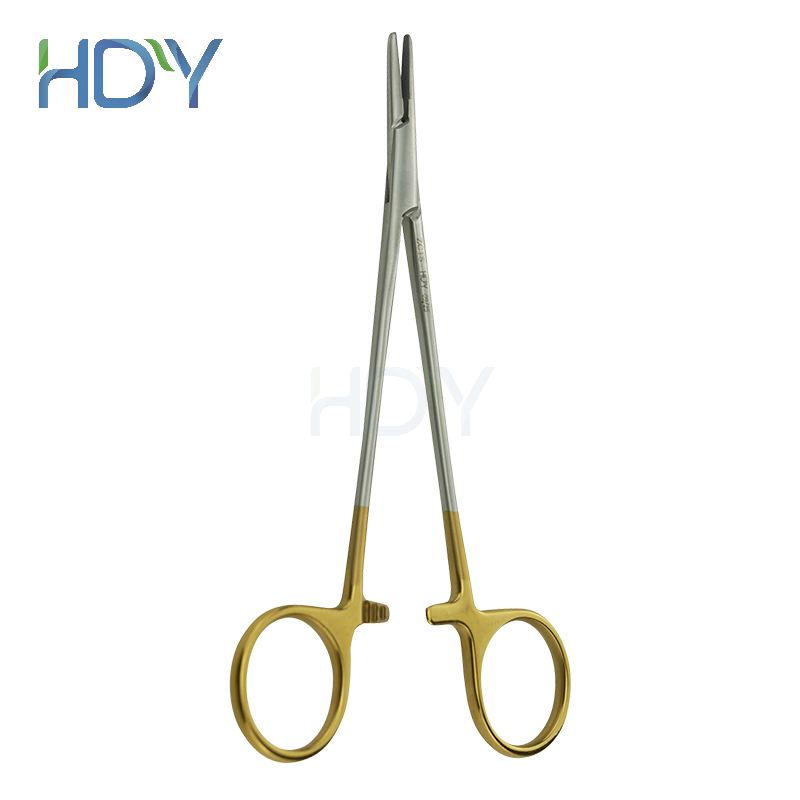
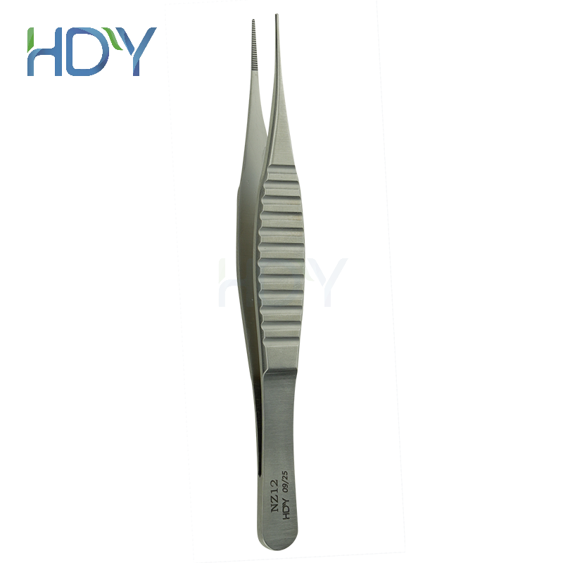
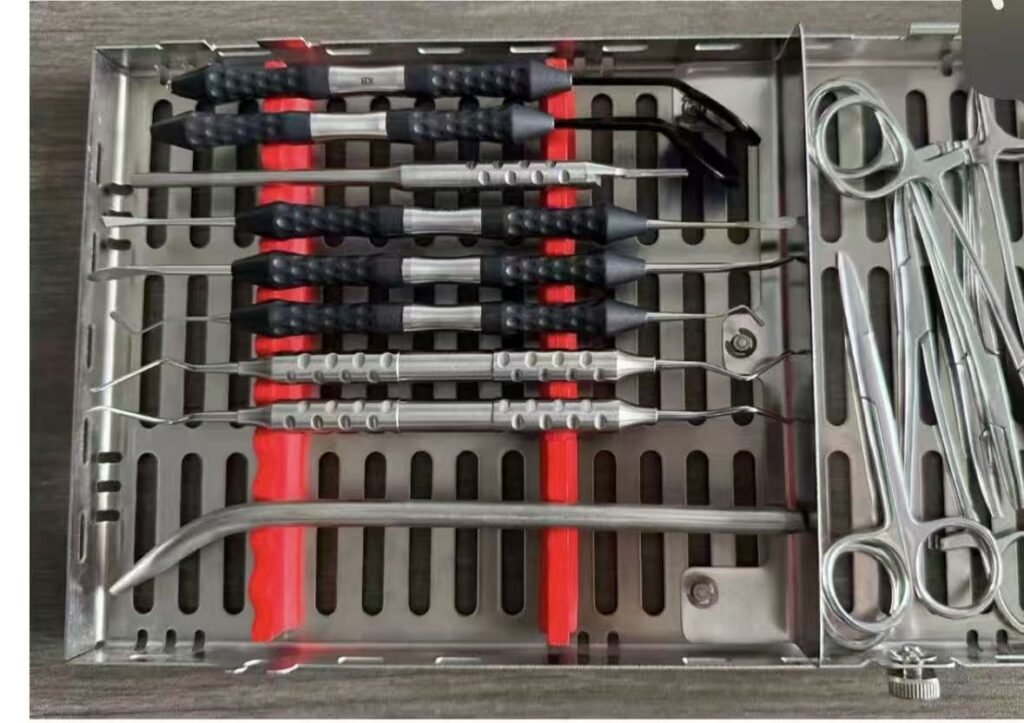
HDY's surgical instrument portfolio includes needle holders, tissue forceps, hemostats and surgical instruments that support controlled tissue handling and wound closure.
Identify the Source
Assess the operative field and determine the likely bleeding source before selecting additional measures.
Apply Local Control
Use appropriate local compression or other clinician-selected hemostatic techniques.
Close When Indicated
Needle holders and suitable sutures can support controlled flap repositioning and wound closure when clinically indicated.
الـ Three Nevers of Prevention
Inferior alveolar and lingual nerve injuries are important complications in mandibular surgery. Prevention depends on anatomical assessment, controlled instrumentation and appropriate surgical visibility.
Avoid uncontrolled apical force
Instrument movement should remain controlled and consistent with the anatomy and intended path of elevation.
Avoid blind lingual instrumentation
Mandibular lingual anatomy requires careful assessment and controlled instrument placement.
Respect the cutting depth
Root separation and bone-related procedures require visibility and controlled depth to protect adjacent structures.
Instrument geometry can support controlled handling, but no instrument can independently eliminate clinical risk. Appropriate case selection, imaging, anatomy knowledge, training and technique remain essential.
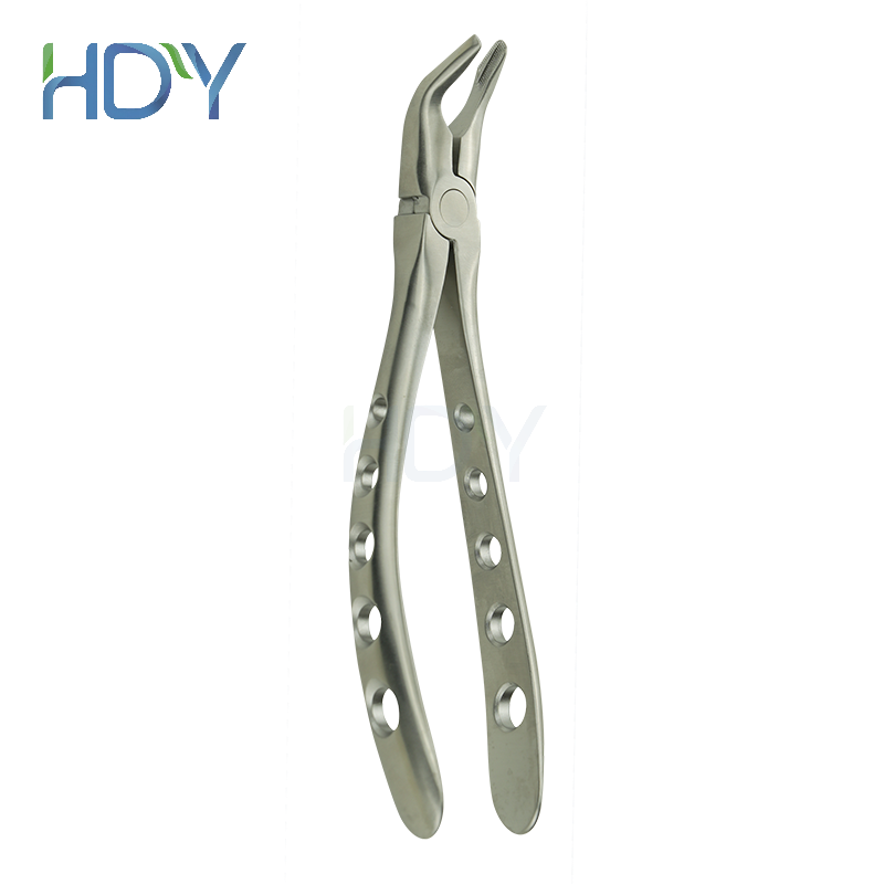
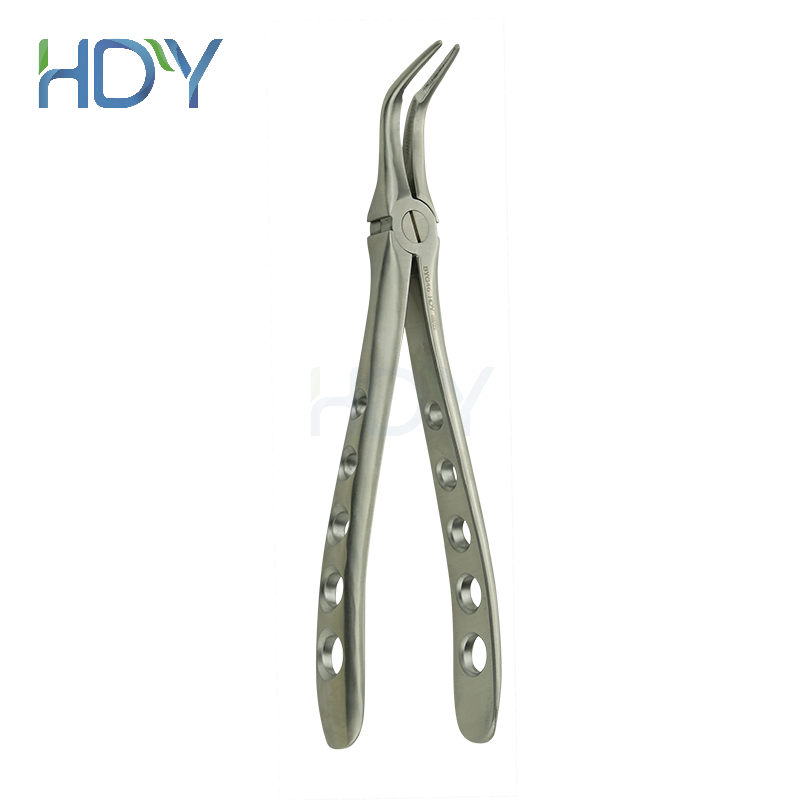
HDY's extraction portfolio includes forceps and root-specific instruments designed for different access and engagement requirements.
Protect the Clot. Respect the Anatomy.
Post-extraction management should focus on protecting the developing blood clot and providing clear postoperative instructions.
Maxillary extraction planning also requires attention to the anatomical relationship between tooth roots and the maxillary sinus. Appropriate radiographic assessment can help identify relevant anatomical relationships before treatment.
Prevention starts before extraction: assess anatomy, review radiographs, select the appropriate instrument and establish a postoperative management plan.
Wrong Tooth Extraction: Systems Matter
Wrong-tooth extraction is a preventable patient-safety incident. The key issue is not simply instrument technique, but verification before the procedure begins.
A structured verification process can include confirmation of the tooth number in the clinical record, review of radiographic information and verbal confirmation between the operator and assistant.
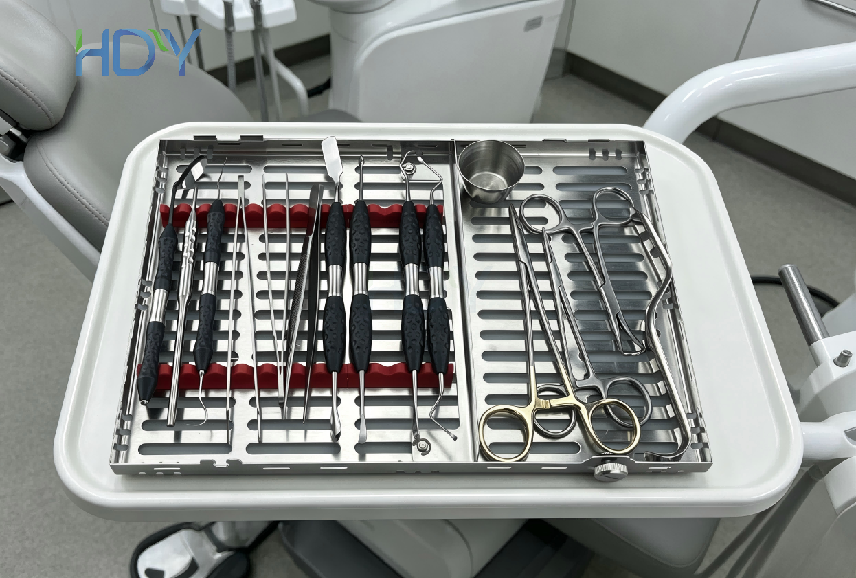
Instrument organization can provide an additional layer of workflow control, particularly when procedure-specific instruments are arranged consistently before treatment.
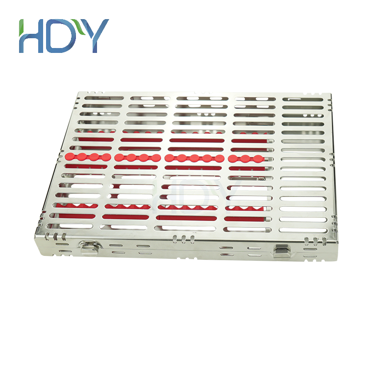
Build a More Structured Extraction Setup
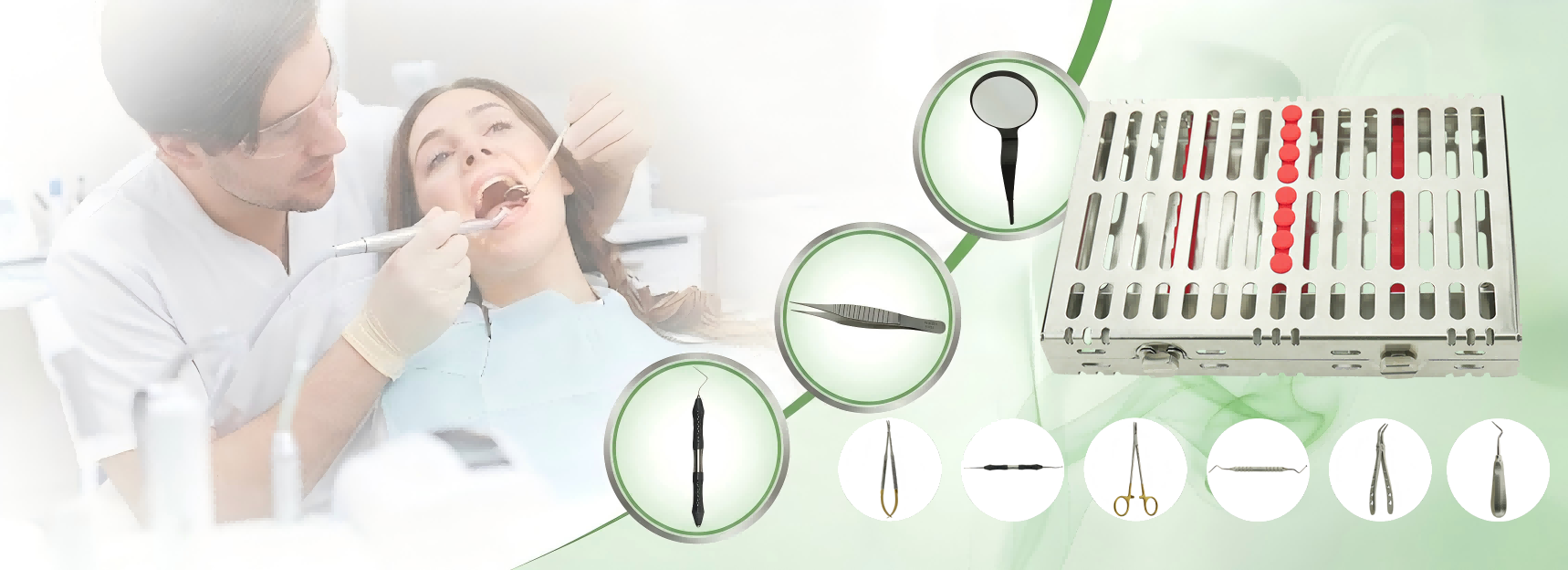
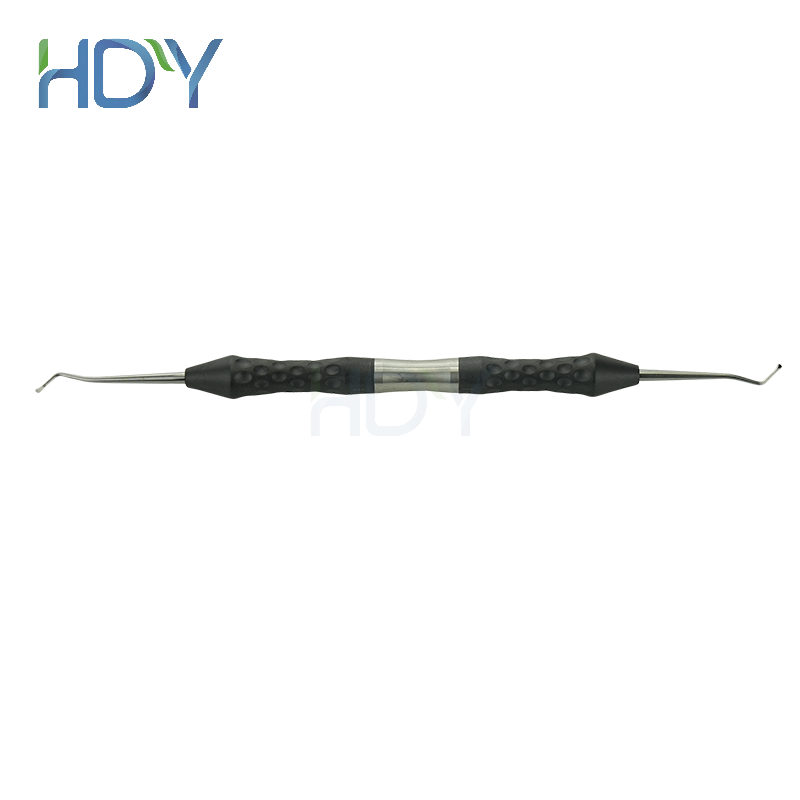
HDY provides a broad range of dental surgical instruments that can support different stages of oral surgery and extraction workflows. Instrument selection should always be based on the clinical case, anatomy, access and the treating clinician's judgment.
ملقط الاستخراج
Tooth-specific and apical forceps for controlled tooth and root engagement.
Explore Extraction Forceps →
ملقط الجذر
Narrow working-end geometry for appropriate root engagement and access.
Explore Root Forceps →
ملقط الأنسجة
Designed for controlled tissue handling during oral surgical procedures.
Explore Tissue Forceps →
حاملات الإبر
Precision needle control for oral surgical suturing and wound closure.
Explore Needle Holders →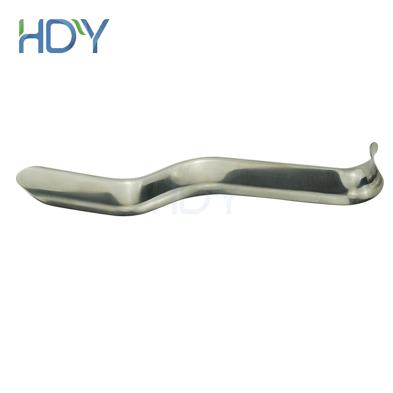

 العربية
 English
 简体中文
 Español
 Deutsch
 Français