

HDY Clinical Instrument Guide
Assess First. Control Pain. Extract Conservatively.
A standardized pathway for routine forceps extraction—from medical and radiographic assessment to tooth-specific forceps selection, controlled socket expansion and root-by-root delivery when indicated.
Clinical principle
Tooth extraction is a surgical procedure—not “grip and pull”
Predictable routine extraction begins before an instrument touches the tooth. The clinician evaluates the patient, establishes profound anesthesia, selects a forceps pattern that matches the tooth and access, and uses controlled movement to expand the socket. Force should remain deliberate, progressive and anatomically directed.
Assess
Review medical history, medications, radiographs, root form, surrounding anatomy, mouth opening and the amount of sound tooth structure available for engagement.
Anesthetize
Confirm adequate local anesthesia before luxation. Technique, agent, dose and supplementary injection must be individualized by a qualified clinician.
Preserve
Use the least traumatic approach consistent with safe delivery. Reassess resistance and section multirooted teeth when an en bloc approach would require excessive force.
Stage 01 · Preoperative assessment
Start with the patient, then return to the tooth
A comprehensive medical and dental history should guide treatment planning. Cardiovascular, respiratory, cerebrovascular, endocrine, renal, hepatic and hematologic conditions may alter timing, setting, hemostatic planning, drug selection or the need for medical consultation. They should not be reduced to a single universal rule.
Radiographs help the dentist assess root number and curvature, periodontal ligament space, bone pattern and proximity to the maxillary sinus or mandibular canal. Restricted opening, limited posterior access and extensive coronal breakdown can change both the instrument and the surgical plan.
- Confirm the indication, tooth identity and consent.
- Review medical conditions, allergies and current medicines.
- Evaluate crown integrity, root anatomy and adjacent structures.
- Plan hemostasis and postoperative instructions before starting.
Stage 02 · Pain control
Do not begin luxation until anesthesia is clinically adequate
Comfort depends on more than the patient reporting “numbness.” The clinician must select an appropriate local-anesthetic technique, inject slowly and confirm pulpal and soft-tissue anesthesia in the intended field. Mandibular molars, acute inflammation and anatomical variation may require a nerve block or supplemental technique. Aspiration, anatomical awareness, dose limits and patient-specific contraindications remain essential.
Reduce injection discomfort
Use an appropriate needle, consider topical anesthetic, enter mobile mucosa where indicated and deposit solution slowly.
Allow onset time
Wait for the selected agent and technique to take effect, then test the operative field before applying extraction force.
Reassess, do not overpower
If the patient experiences pain, stop and reassess anesthesia. More mechanical force is not a substitute for adequate pain control.
Supporting instruments · Examine, separate, then engage
HDY instruments used before forceps delivery
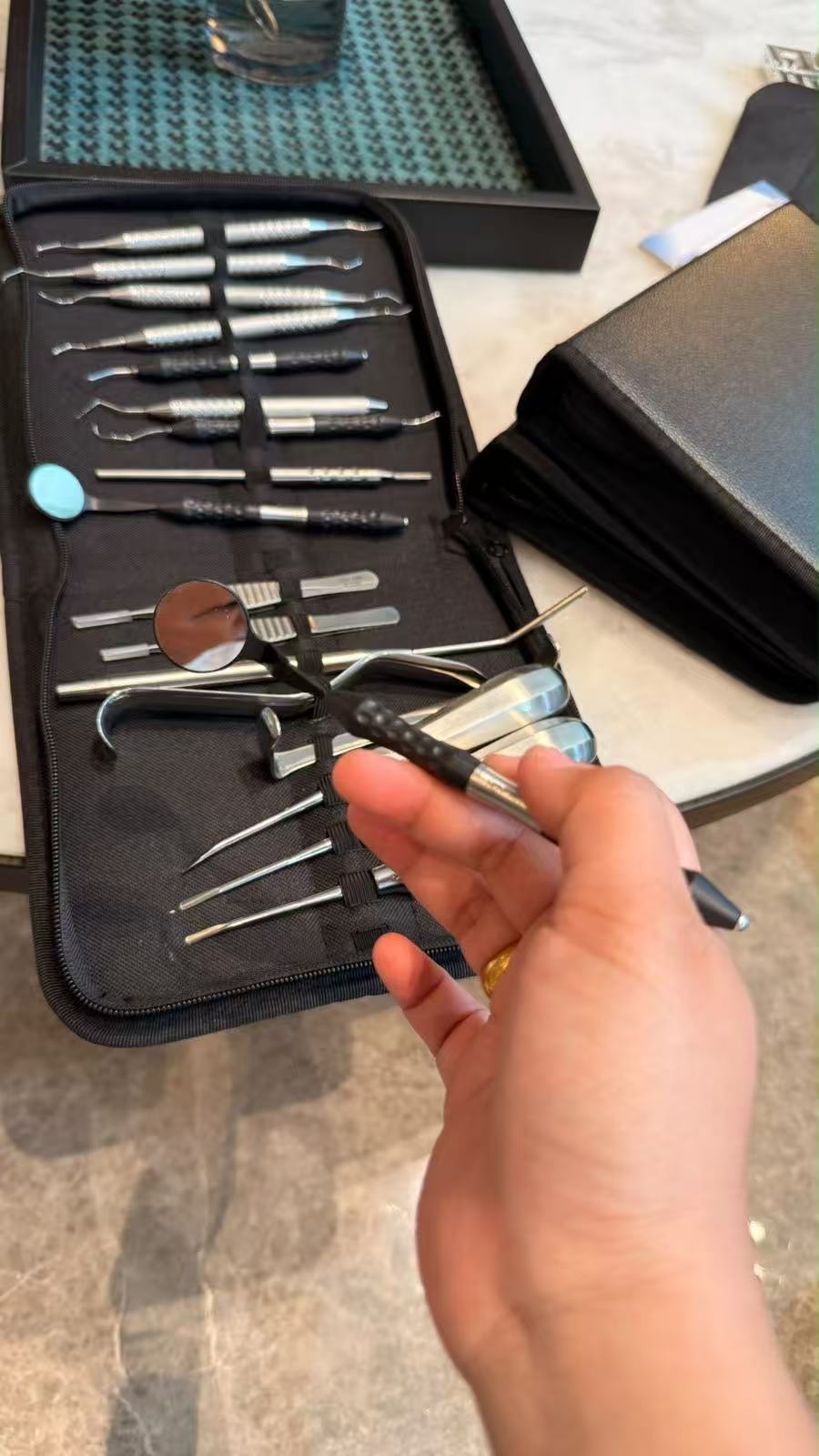
Once adequate anesthesia has been confirmed, fine diagnostic and periodontal-ligament instruments can support a more controlled transition to forceps extraction. The explorer assists visual and tactile assessment, while anterior- and posterior-specific periotomes are used to sever periodontal-ligament fibers where clinically indicated. These instruments prepare access; they are not substitutes for diagnosis, anesthesia or controlled forceps technique.
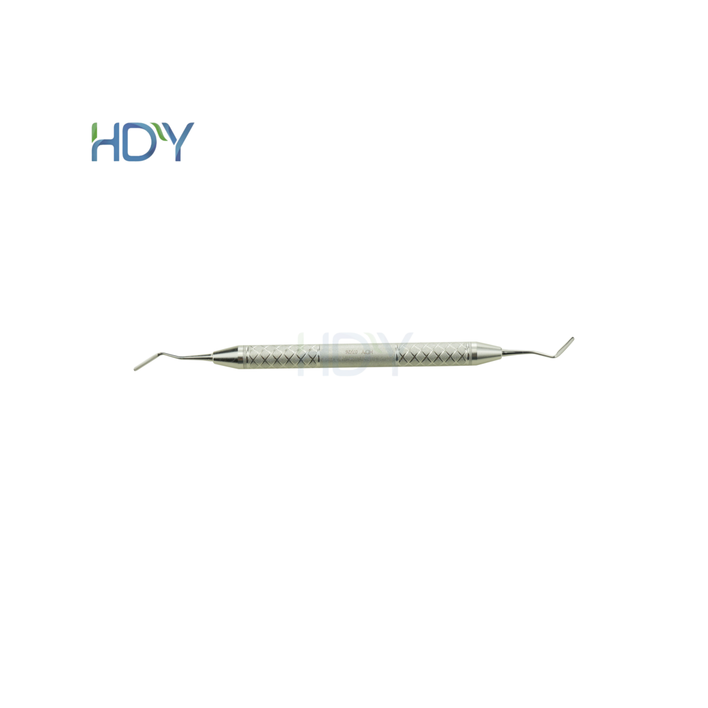
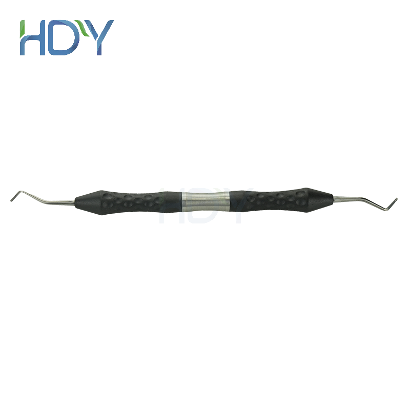
Posterior working-end geometry for controlled periodontal-ligament separation where access and anatomy permit. View BY05G →

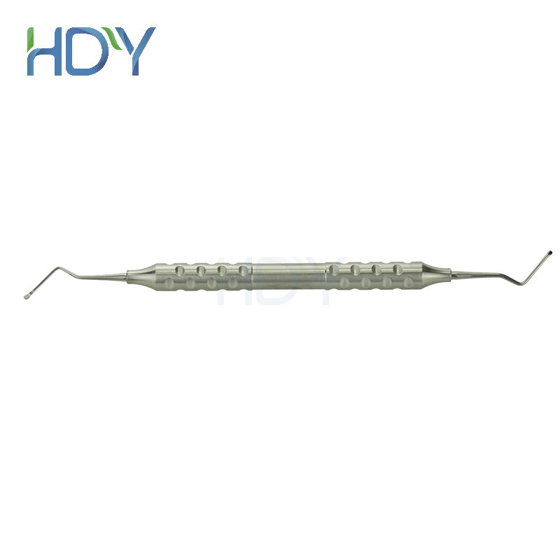
Anterior blade design for controlled periodontal-ligament separation around front teeth. View BY06G →
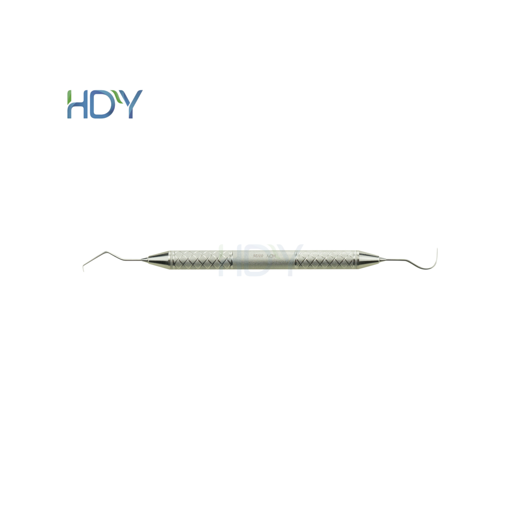
A fine diagnostic explorer for controlled tactile examination and confirmation of accessible margins. The website model is listed as TZ5G. View TZ5G →
Stage 03 · Forceps selection
Fit the beaks to the cervical anatomy and align with the long axis
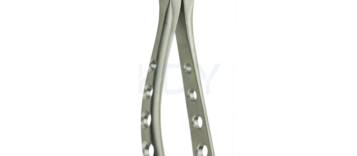
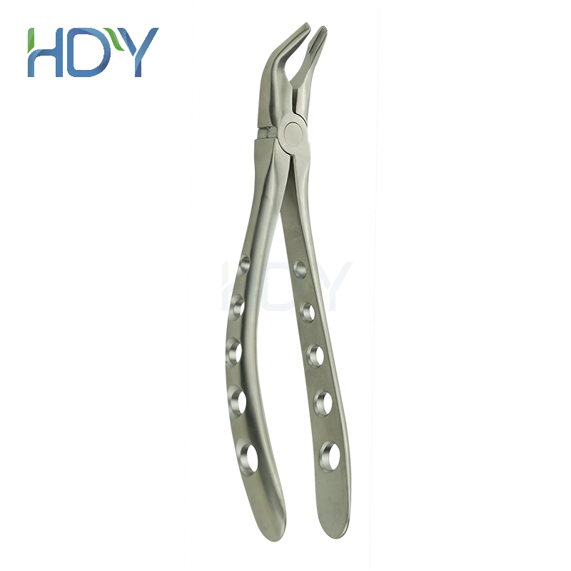
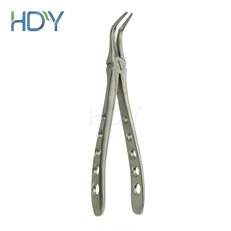
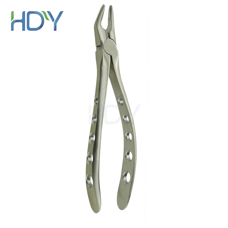
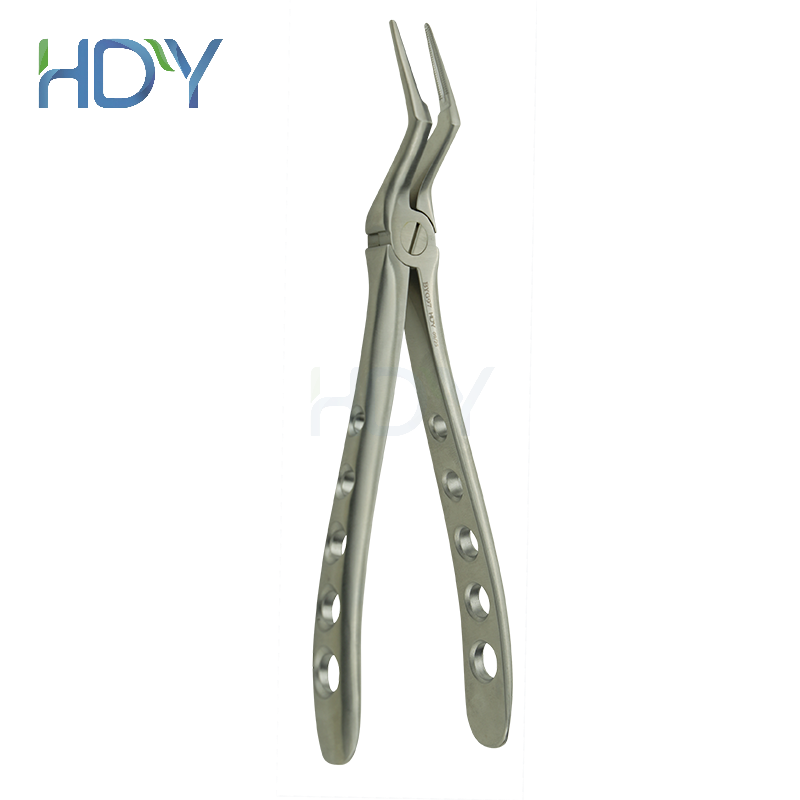
When sufficient crown or root structure can be engaged safely, extraction forceps can support controlled delivery. Beaks should seat as apically as the clinical situation permits, engage stable tooth structure and align with the intended path of movement. Pattern selection should reflect tooth location, root form and access.
| Clinical area | Typical forceps geometry | Controlled movement | Planning note |
|---|---|---|---|
| Maxillary anterior teeth | Straight or narrow beaks | Buccopalatal movement; limited rotation for suitable single conical roots | Match rotational force to root shape and curvature. |
| Maxillary premolars | Narrow beaks adapted to cervical contour | Measured buccopalatal expansion | Root bifurcation or thin roots may increase fracture risk. |
| Maxillary molars | Right- and left-specific beaks adapted to buccal furcation and palatal root | Controlled buccopalatal movement | Confirm side and furcation engagement before loading. |
| Mandibular anterior teeth | Narrow beaks with handle-to-beak angulation for access | Small buccolingual movements | Avoid indiscriminate rotation of flattened or curved roots. |
| Mandibular molars | Molar or cowhorn-pattern beaks, when appropriate | Progressive buccolingual expansion | Section roots when anatomy or resistance makes en bloc delivery traumatic. |
| Retained roots | Long, narrow apical/root forceps | Precise engagement with minimal uncontrolled leverage | Escalate to a surgical approach when visibility or purchase is inadequate. |
Stage 04 · Controlled movement
Expand the socket progressively—do not yank the root
- Release the gingival attachment where indicated and establish clear access.
- Seat the beaks apically against stable cervical tooth structure.
- Support the alveolus and protect adjacent soft tissue with the non-dominant hand.
- Apply slow, small-amplitude movement toward the thinner cortical plate, then in the opposite direction.
- Increase the range only as the socket expands; use rotation only when root anatomy supports it.
- Deliver along the path of least resistance and inspect the tooth or roots after removal.
Stage 05 · Multirooted teeth
Separate roots when whole-tooth delivery would demand excessive force
A heavily carious multirooted molar, divergent roots, dense supporting bone or limited coronal purchase can make en bloc extraction unpredictable. When clinically indicated, sectioning the crown and roots converts one complex path of removal into smaller, more controllable paths. Each root can then be mobilized and delivered with an instrument suited to its shape and available access.
Sectioning is a surgical decision, not a universal step. The clinician must maintain visibility, protect adjacent structures, control the bur and irrigation, and confirm complete removal or document any intentionally retained fragment according to accepted clinical guidance.
HDY instrument engineering
Working-end geometry, joint stability and controlled handling
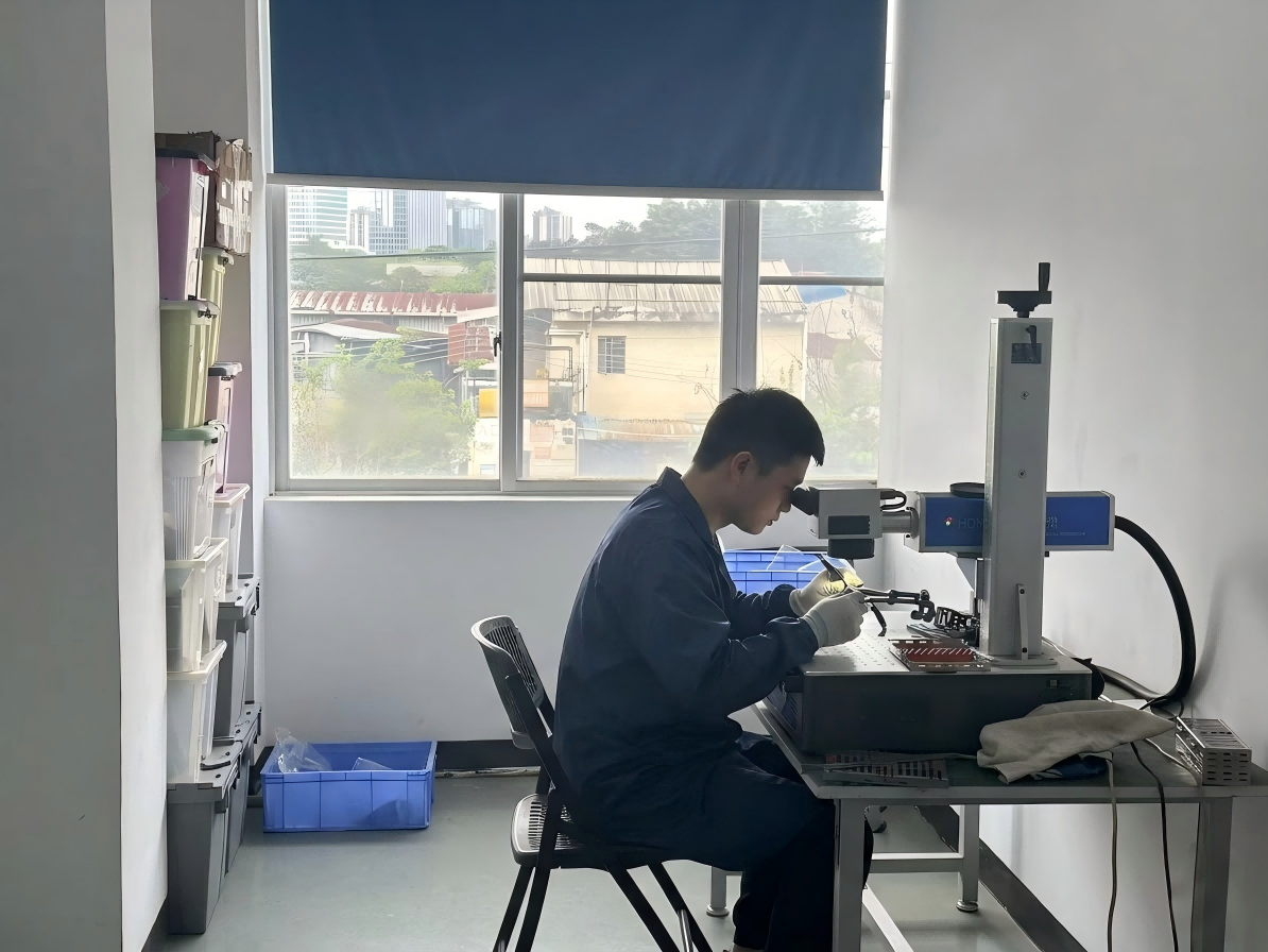
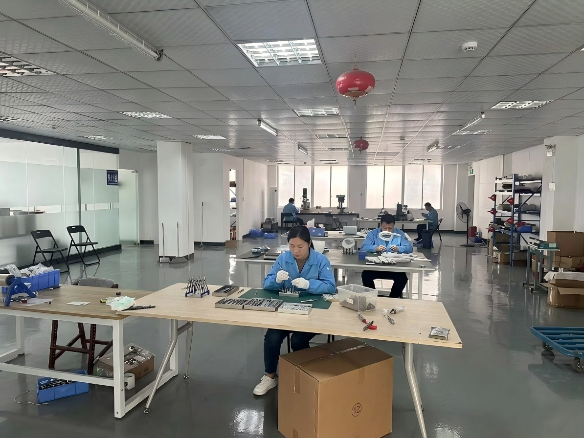
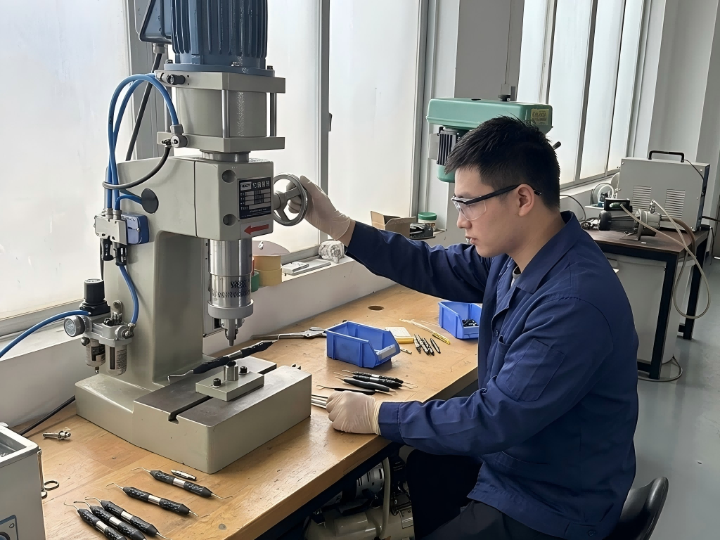
HDY organizes extraction forceps by clinical access and intended engagement. Selected forceps may use near-net-shape metal injection molding (MIM) for repeatable component geometry, followed by machining, finishing and inspection as required by the product specification. Serrated beaks, apical patterns and root-specific working ends are designed to provide controlled purchase when correctly matched to the case.
Product-specific material grade, dimensional tolerances, sterilization parameters and regulatory status should be confirmed from the applicable technical file or quotation. Instrument design can support control, but it does not replace diagnosis, training or sound surgical technique.
Frequently asked questions
Routine dental extraction forceps FAQ
How should a dentist select extraction forceps?
Selection should reflect tooth location, crown and root anatomy, access, furcation form, remaining tooth structure, radiographic findings and the planned surgical approach. A “universal” pattern is useful only when its geometry truly matches the case.
When should a multirooted tooth be sectioned?
Sectioning may be appropriate when root divergence, limited remaining crown, dense bone or fracture risk makes en bloc removal unnecessarily traumatic. The treating clinician decides after clinical and radiographic assessment.
Are apical forceps the same as routine extraction forceps?
No. Apical or root forceps have narrower working ends intended for retained roots or limited subgingival access. They should be selected only when their design provides safe visibility and purchase.
Can a forceps design guarantee a minimally invasive extraction?
No instrument can guarantee an outcome. Appropriate case selection, anesthesia, anatomical knowledge, controlled technique and timely conversion to a surgical approach are equally important.
Selected clinical references
Build a tooth-specific extraction instrument set
Contact HDY for forceps specifications, distributor pricing, samples, private-label packaging or OEM and ODM dental instrument development.

 English
  简体中文
简体中文  Español
  Deutsch
  Français